Prevalence of otomycosis in Constantine, Algeria: A cross-sectional study of two months

Article Sidebar
-
Otomycosis, Antifungal, Filamentous Fungi, Yeast Fungi, Candida, Aspergillus, Fungal Susceptibility, Fungal Otitis
Abstract
Otomycosis is a fungal infection of the external auditory canal. Over a two-month period 34 ear samples were analyzed in mycology laboratory at the Military Hospital Cdt Abdelali Benbaatouche, of Constantine, Algeria.
Our study revealed that the positivity rate of otomycosis is 59%, with a male predominance (79%). The age group with the highest prevalence of otomycosis was the 35-49 years-old.
The occurrence of otomycosis was statistically more common in patients using cleaning cotton bud (42.9%). The left ears were the most affected (57%). Otalgia was the most common presenting symptom (35.7%).
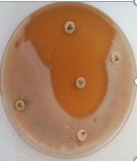
Mycological study revealed yeasts are the most dominant (57%) compared to filamentous fungi (43%). The predominant species was A. niger (43%), and four Candida genus species were found, C. albicans (22%), C. parapsilosis and C. dubliniensis with isolation percentage of 14% respectively, followed by C. tropicalis (7%).
- dubliniensis and C. albicans were resistant to amphotericin B, griseofulvin, and nystatin. However, they were susceptible to ketoconazole, clotrimazole, miconazole, and econazole.
Full text article
References
1.Aboulmakarim, S., Tligui, H., El Mrini, M., Zakaria, I., Handour, N., & Agoumi, A. (2010).
Otomycoses: étude clinique et mycologique de 70 cas. Journal de Mycologie Médicale, 20(1),
-52.
2.Lecanu J.-B., Erminy M., Faulcon P., Théoleyre B. 2008. Otomycose. EMC (Elsevier Masson
SAS, Paris), Oto-rhino-laryngologie, 20-080-A-10
3.Munguia, R., & Daniel, S. J. (2008). Ototopical antifungals and otomycosis: a review.
International journal of pediatric otorhinolaryngology, 72(4), 453-459
Gray, R. F., Sharma, A., & Vowler, S. L. (2005). Relative humidity of the external auditory
canal in normal and abnormal ears, and its pathogenic effect. Clinical otolaryngology, 30(2),
-111
Y Merad, Z Belmokhtar, A Messafeur, M Belkacemi, MFM Réda. Prevalence of superficial mycosis in breast cancer patients: a Cross-Sectional Study. Cancer Sci Res 2 (4), 1-4, 2019
Y Merad, H Adjmi-Hamoudi, K Lahmer, E Saadaoui, S Cassaing, A berry. Les otomycoses chez les porteurs d’aides auditives: étude rétrospective de 2010 à 2015. Journal de Mycologie Médicale, 2016
Y Merad, H Adjmi-Hamoudi, FZS Merad, S Djeriou. Otomycosis in the fisherman community: A survey at Bénisaf harbour, Aïn Témouchent, Algeria. J. Med. Biomed. Appl. Sci 6, 118-120, 2018
Y Merad, H Derrar, M Belkacemi, A Drici, Z Belmokhtar, AEM Drici. Candida albicans mastitis in breastfeeding woman: an under recognized diagnosis.Cureus 12 (12), 2020
Y Merad, L Thorayya, M Belkacemi, F Benlazar. Tinea unguium, tinea cruris and tinea corporis caused by Trichophyton rubrum in HIV patient: a case report. J Clin Cases Rep 4 (4), 89-92, 2021
M Yassine, AH Haiet. Self-injury in schizophrenia as predisposing factor for otomycosis. Medical mycology case reports 21, 52, 2018
Y Merad, AA Moulay, H Derrar, M Belkacemi, NA Larbi-Cherak. Aspergillus flavus onychomycosis in the right fourth fingernail related to phalynx fracture and traumatic inoculation of plants: A vegetable vendor case report. Am. Res. J. Dermatol 2, 1-4, 2020
Bhally, H. S., Shields, C., Lin, S. Y., & Merz, W. G. (2004). Otitis caused by Scedosporium
apiospermum in an immunocompetent child. International journal of pediatric
otorhinolaryngology, 68(7), 975-978
K.D. Adoubryn, V.K. N’Gattia, G.C. Kouadio-Yapo, L. Nigué, D.K. Zika, J. Ouhon. Epidemiology of otomycoses at the University Hospital of Yopougon (Abidjan-Ivory Coast). Journal de Mycologie Médicale. Volume 24, Issue 2
Kurnatowski, P., & Filipiak, A. (2001). Otomycosis: prevalence, clinical symptoms, therapeutic
procedure. Mycoses, 44(11‐12), 472-479.
Ozcan, K. M., Ozcan, M., Karaarslan, A., & Karaarslan, F. (2003). Otomycosis in Turkey:
predisposing factors, aetiology and therapy. The Journal of Laryngology & Otology, 117(1),
-42
Dorko E, Jenca A, Orencák M, Virágová S, Pilipcinec E. Otomycoses of candidal origin in eastern Slovakia. Folia Microbiol (Praha). 2004;49(5):601-4.
Cheriet N et al. Contribution à l’étude de la situation épidémiologique des otitis d’origines fongiques. (2017), these
Ali K, Hamed MA, Hassan H, Esmail A, Sheneef A. Identification of Fungal Pathogens in Otomycosis and Their Drug Sensitivity: Our Experience. Int Arch Otorhinolaryngol. 2018 Oct;22(4):400-403.
Kurnatowski, P., & Filipiak, A. (2001). Otomycosis: prevalence, clinical symptoms, therapeutic
procedure. Mycoses, 44(11‐12), 472-479.
Pontes ZB, Silva AD, Lima Ede O, Guerra Mde H, Oliveira NM, Carvalho Mde F, Guerra FS. Otomycosis: a retrospective study. Braz J Otorhinolaryngol. 2009 May-Jun;75(3):367-70.
Fasunla J, Ibekwe T, Onakoya P. Otomycosis in western Nigeria. Mycoses. 2008 Jan;51(1):67-70.
Authors
Copyright (c) 2024 Journal of Current Medical Research and Opinion

This work is licensed under a Creative Commons Attribution 4.0 International License.