Ultrasound studies on Mycoplasma bronchopneumonia

Article Sidebar
Abstract
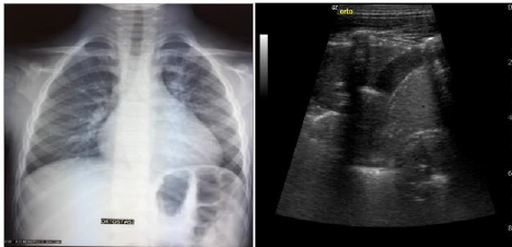
Backround Pediatric bronchopneumonia represents a clinical challenge, especially when it comes to the identification of its etiology. Working Hypothesis We performed a retrospective study on 100 patients admitted to our Pediatric Department. Only patients with bronchopneumonic thickening were selected, discharged with a diagnosis of Community - Acquired Pneumonia (CAP) or bronchopneumonia. The purpose of our study was to identify Mycoplasma Pneumonia based on lung ultrasound (LUS) findings. Methodology At least two lung LUS were performed on each patient: on admission and few days after start of therapy, with most patients undergoing a third ultrasound evaluation approximately one week after discharge. These reports were collected for each patient together with clinical and laboratory data. The study population was divided into two groups: patients who tested positive for Mycoplasma pneumoniae (Myc-CAP) and negative ones (non-Myc-CAP). All patients performed serological test for determination of anti-mycoplasma antibodies, and in doubtful cases also molecular test with PCR on pharyngeal exudate. Results The results obtained after statistical analysis showed no significant differences in LUS findings between the two groups, that could allow a positive differential diagnosis of Myc-CAP without resorting to laboratory testing. Conclusions LUS undoubtedly represents a valid and irreplaceable help in the morphological study of pulmonary lesions over the course of disease from the time of admission to follow-up.
Full text article
Generated from XML file
Authors
Clelia, T., Marella, P. ., Onofrio, I., Vincenzo, B. ., Teresa, D. B. ., Maria, F. ., Francesca, L. ., Egisto, S. ., Maria, S. ., & Ignazio, L. . (2022). Ultrasound studies on Mycoplasma bronchopneumonia. Journal of Current Medical Research and Opinion, 5(08), 1301−1315. https://doi.org/10.52845/CMRO/2022/5-8-6
Copyright and license info is not available